Knee & Sports Injury Clinic
Bristol
Email: boc@orthopaedics.co.uk


Medical Information
Explore detailed information about a range of joint problems and treatments, including medications, surgery, physiotherapy and rehabilitation. Reading this will help you understand more about your own condition. There is also a glossary with explanations of many medical terms used in orthopaedics. You can find out even more by following the links page to other related websites, journals or professional medical associations.
Hover over links below to view summary or click on the link to view full article:
Total Knee Replacement: A Patient’s Guide
Author: DAVID P JOHNSON MB ChB FRCS FRCS. MD
Consultant Orthopaedic Surgeon
THE ANATOMY OF THE KNEE
The knee comprises the joint between the femur and the tibia but also the joint between the patella and the front of the femur. Either or all of these parts of the knee may be affected by arthritis to various degrees. A total knee replacement replaces the surfaces of the knee with plastic and metal components. The femoral replacement is a smooth metal component which fits snugly over the end of the bone. The tibial replacement is in two parts, a metal base plate sitting on the bone and a plastic insert which sits between the metal base on the tibial and the femoral component. If necessary the patella surface (under the knee cap) is replaced with a plastic button which glides over the metal surface of the femoral replacement. However, the patella is occasionally satisfactory and may not require replacement.
The components are usually cemented to the bones in order to secure fixation. In certain circumstances special components may be “press fitted” to the bones without the additional use of cement. These components use micro-porous metallic surfaces and may have an additional hydroxyapatite coating to promote osteo-integration or bonding to the bone. These techniques may be considered and appropriate for younger patients.
EXERCISES AND MEDICATION : PRIOR TO SURGERY
Prior to surgery and post operatively it is important to strengthen the muscles of the leg and to reduce the stiffness. Regular exercises should be undertaken to do this. Static quadriceps exercises consist of tensing the muscle on the front of the thigh whilst the knee is straight. Hold the contraction for 5 to 10 seconds, rest for 5 or 10 seconds and begin again. This should be repeated 10-50 times. Whilst lying on your back the straight leg should be lifted into the air and held for 5 to 10 seconds, then lowered rested for 5 to 10 seconds and repeated 10 to 50 times. Sitting on a high chair or table and bending the knee over the edge will improve knee bending. The good leg may be crossed over the bad one to assist.
TESTS AND SCANS
Prior to admission to hospital blood tests, a urine test, a knee X-ray, a chest X-ray and an ECG or heart tracing will be performed. Blood will be cross-matched so as to be available for transfusion following surgery. If you wish to arrange for your own blood to be pre-donated and ready for autologous transfusion, this should be done some weeks in advance and will require a trip to the blood bank on several occasions. It is a routine to use blood salvage techniques during and following surgery, which minimises the need for blood transfusions. Minimally access techniques also reduce the need for blood transfusions. Prior to the operation any tablets or medications you take, or allergies you may have to medications, should be brought to the attention of the surgeon. You should stop anti-inflammatory arthritis tablets for one week prior to surgery. Take only Paracetamol for pain relief during this period. Please notify your surgeon and anaesthetist in advance if you are taking any anti-coagulants (blood thinners), hormone replacement tablets, the Pill or suffer from diabetes or any other significant medical condition.
SURGICAL TECHNIQUE
A general anaesthetic is generally used, sometimes a spinal injection is preferred. To be able to completely replace the surface of the knee joint a 15cm incision is made down the front of the knee and the joint is opened. If a semi or partial knee replacement is being performed, a smaller incision sometimes of only 5 cm in length may be used.
MINIMALLY INVASIVE SURGERY
Minimally invasive surgery involves the use of smaller wound incisions and special instrumentation to enable surgery to be undertaken. These techniques usually result in significant advantages in respect to improve the speed of recovery, speed of mobilization, shorten hospital stay reduce the period off work and reduce the time until functional and sporting activities can be resumed. The techniques also usually reduce the amount of post-operative pain experienced and the need for post-operative pain relief and analgesia. In knee replacement these techniques are applicable and routinely used by your surgeon in uni-compartmental knee replacement and increasingly in Total Knee Arthroplasty.
Once the wound has been developed and the knee exposed. The bony overgrowth, which commonly occurs in arthritis of the knee, is trimmed away and the joint surfaces removed. This involves some shaping of the bone so that the joint replacement components sit firmly on the bone. The components will either be fixed in situ with acrylic cement or for young patients special components with a roughened or porous surface will be used. The bone then grows into the roughened surfaces anchoring it.
Once the total knee replacement has been inserted the knee joint is closed over drainage tubes. These tubes take away the bleeding from the knee. They stay in the knee overnight. The knee will have a dressing afterwards and be bandaged. You will have a drip to administer fluids whilst you do not feel like eating or drinking. A blood transfusion may be given if required. Initially the knee may be painful. Powerful pain-killing tablets and injections will be prescribed. It is usual for these to be required regularly for 1 or 2 days and for 1 to 2 weeks intermittently, so do not be afraid to ask if you are in pain. Further blood tests and X-rays will be taken. Injections or tablets to thin the blood and to prevent thrombosis will be given.
The recovery from the operation requires about 3-7 days in hospital. In this time physiotherapy is commenced. Initially the leg will be placed on a continuous passive motion machine to gently move the knee through a pre-set range of motion. In addition, exercises to improve the strength of the quadriceps muscles are performed.
WOUND DRESSING AND SUTURES
The crepe bandage, which is applied in theatre, may be removed after 4 days if the wound is satisfactory. The general practitioner or practice nurse should remove the sutures after 14 days. Sometimes arrangements are made for patients to return directly to the hospital for this..
MEDICATION
If the joint replaced was the only joint affected by arthritis, no further anti-inflammatory tablets will be required once the initial post-operative discomfort settles. If other joints are affected or you suffer from rheumatoid arthritis, the tablets may be restarted, if possible after an interval of 2 to 3 weeks. Pain killers may be taken during this time before anti-inflammatory tablets are restarted.
PHYSIOTHERAPY
The day following surgery the physiotherapist will get you out of bed to commence walking with the help of crutches or a walking frame. The machine will be removed increasingly for you to perform your quadriceps exercises. After the first few days the range of motion pre-set on the continuous passive motion machine will be increased. The physiotherapist will also begin to encourage you to bend the knee. Sitting over the side of the bed may assist. The early exercises and mobilising of the knee will cause some discomfort and swelling. However, this is normal and is just the healing process occurring. Any swelling or discomfort in the calf muscle should be brought to the attention of the nursing staff or after discharge, to your general practitioner.
After 5-10 days usually you are able to walk with minimal or no pain, with the assistance of sticks, crutches or a frame. You should be able to manage stairs with the assistance of a banister, and to care for yourself around the home. When this is possible you will be discharged from hospital. Whilst at home the exercise program of quadriceps exercises should be vigorously continued. Approximately 10 minutes each hour will be ample. Out patient physiotherapy sessions should be arranged during this period. Resisted exercises and training may be undertaken if desired after 6 weeks; this would be beneficial. The progress is a little slow at this stage. If the knee becomes more swollen or more painful than during the first day, please return to your general practitioner for early review. Prior to discharge from hospital the ward staff on behalf of your surgeon will usually provide an appointment for review after 4-6 weeks.
RETURN TO WORK AND SPORT
If your job is sedentary and mostly sitting you may wish to return after only 3 – 6 weeks. If your job is physically demanding and requires standing or walking for most of the day, your return to work may take several months. Driving can usually be performed after 4-6 weeks providing that the knee is pain free and you are able to control the car with foot pedals and make an emergency stop.
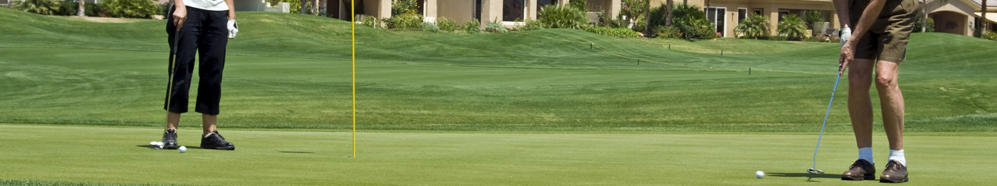
Resisted exercises and training may be undertaken. This will speed up the rehabilitation and should be performed prior to undertaking more vigorous sports. Swimming is often possible after 3 weeks. Return to golf, gentle tennis or badminton may take 3 months. Jogging and squash is not advised.
Please visit www.orthopaedics.co.uk for more information.
DAVID P JOHNSON
MB ChB FRCS FRCS. MD
Spire Glen Hospital, Redland Hill, Bristol BS6 6HW. UK
E-Mail: boc@orthopaedics.co.uk
Web site: www.orthopaedics.co.uk
Full text pdf
© OrthopaedicsOpinionOnline 2011 www.OrthopaedicOpinionOnline.co.uk
Disclaimer: The views expressed in this article are not necessarily those of Orthopaedic Opinion Online or the author. The information is provided for general background reading only and should not be relied upon for treatment. Advice should always be taken from a registered medical practitioner for individual circumstances and for treatment of any patient in any circumstances. No liability is accepted by Orthopaedic Opinion Online, or the author in respect to the information provided in respect of the content or omission or for any reason or as a result of treatment in individual circumstances. This information is not for use in the USA.