Knee & Sports Injury Clinic
Bristol
Email: boc@orthopaedics.co.uk

Treatments
Browse through a rich range of information to find details of various treatments available for your condition. These include details on the treatment options, medications, physiotherapy and injections. Details are also provided on the range of surgical procedures and the rehabilitation following surgery.
You can also find out more about how to access private treatment when paying for yourself and the statutory quality criteria and success rates of our treatments.
Hover over links below to view summary or click on the link to view full article:
Anterior Cruciate Ligament Reconstruction: Patellar Tendon Graft
THE ANATOMY OF THE KNEE
The knee comprises the joint between the femur and the tibia but also the joint between the patella and the front of the femur. Between the femur and the tibia sit two crescentic cartilages or menisci. These fibro-cartilaginous discs dissipate the compressive forces between across the knee and thereby avoid excessive loading, wear and damage. The ligaments around the knee stabilise the knee. They include the collateral ligaments; medial and lateral, lying either side of the knee and the cruciate ligaments, anterior (ACL) and posterior cruciate ligament (PCL), lying within the joint. The fibrous capsule, which surrounds the knee, completes the stability of the joint. The two most important groups of muscles supporting the knee are the quadriceps muscle, which is the large bulk of muscle lying at the front of the thigh, and the hamstrings which lie behind the thigh.
The Anterior Cruciate Ligament (ACL) runs posteriorly and upwards from its insertion on the central anterior position on the tibia to its origin on the posterior lateral wall of the inter-condylar notch of the femur. It is approximately 2.5cm in length and approximately 3000N in strength. The techniques of reconstruction are the “gold standard” patellar tendon technique or the hamstring technique.
The bone-patellar-tendon graft is a strip of bone 10mm in width harvested from the inferior anterior surface of the patella and its attached strip of patellar tendon. The corresponding block of bone from the tibia to which the tendon attaches is also harvested providing a graft 10mm in width of patellar tendon with an attached block of bone at either end. Using arthroscopic techniques this graft is then fed through drill holes across the knee and fixed in situ with screws at either end. Mr. Johnson has for the last 10 years utilized absorbing screws, which naturally dissolve and avoid the need for removal at a later date. Use of this graft does result in some weakness of the quadriceps muscle which usually recovers. Some discomfort on kneeling may also be experienced. This technique is generally regarded as the gold standard and slightly more successful in providing knee stability than the hamstring graft technique.
The hamstring technique uses the semitendinosus and gracilis tendons from the hamstring muscles at the medial side of the knee. In a similar way to the Bone-patellar-tendon graft this is then fed across the knee. Fixation of this graft can be undertaken in a variety of ways. The use of this graft is associated with some hamstring and quadriceps weakness which usually recovers. It is associated with less discomfort on kneeling.
EXERCISES AND MEDICATION PRIOR TO SURGERY
Prior to surgery and post operatively it is important to strengthen the muscles of the leg and to reduce the stiffness. Regular exercises should be undertaken to do this. Static quadriceps exercises consist of tensing the muscle on the front of the thigh whilst the knee is straight. Hold the contraction for 5 to 10 seconds, rest for 5 or 10 seconds and begin again. This should be repeated 10-50 times. Whilst lying on your back the straight leg should be lifted into the air and held for 5 to 10 seconds, then lowered rested for 5 to 10 seconds and repeated 10 to 50 times. If possible these exercises should be undertaken against weight or resistance or in a gym using exercise equipment.
Anti-inflammatory tablets (Indomethacin, Voltarol, Brufen, Naprosyn etc) must be stopped 2 days before surgery. On the morning of surgery patients should fast from midnight and arrive at the hospital at 7.20am. For afternoon surgery you will be fasted after 8am. Prior to the operation any tablets or medications you take, or allergies you may have to medications, should be brought to the attention of the surgeon. Please notify your surgeon and anaesthetist in advance if you are taking any anti-coagulants (blood thinners), hormone replacement tablets, the Pill or suffer from diabetes or any other significant medical condition.
SURGICAL TECHNIQUE
The anaesthetist and Mr. Johnson will see you before surgery. The operation is performed under spinal or general anaesthesia. There will be one or two small incisions in the front of the knee either side of the patellar tendon. Two small screws are usually used to anchor the ligament in the bone tunnels. Mr. Johnson usually currently uses third generation dissolving poly-lactic acid / tri-calcium phosphate screws. These usually dissolve over a period of three years and do not usually require removal. Following the operation patients usually wake up with a brace on the leg and a continuous passive motion machine that is designed to gently move the knee even while you are asleep. This is used to maintain a range of mobility in the joint to speed your recovery. A removable knee splint may be used to support the knee during the recovery period.
The aim of reconstruction of the anterior cruciate ligament (ACL) is to replace the damaged ligament with a graft that reproduces the features and stability provided by the anterior cruciate ligament. The method employed is to take from the patients own body a piece of tendon of similar size and strength to the ACL and fix it within the knee joint in the same position as the old ACL. The use of artificial ligaments is not as successful. The middle third portion of the patella tendon between the kneecap and the tibia with an attached piece of bone at each end is the available structure which results in the best success rate and return to sports (Regeneration occurs at the site from which the graft is taken). Alternately hamstring tendons can be harvested from the muscles at the back of the knee. The technique of ACL reconstruction, developed in 1990, is continually being improved and adjusted based on experience and research. The procedure should be undertaken arthroscopically through keyhole surgery involving two small 6mm holes in the front of the knee. Arthroscopic ACL reconstruction results in less post operative pain, more accurate placement of the graft in the knee, deals with other problems in the knee, allows a more rapid and successful recovery of movement and function.
The arthroscopic technique involves making drill holes through the tibia into the knee joint, and from inside the joint into the femur. The graft is then passed through these holes so that it lies in the correct position through the joint. The graft is then secured at each end with a screw. It should also be emphasized that at the time of arthroscopy additional damage may be found to the cartilages or joint surface. An additional procedure may be undertaken to correct this and this can usually be done simultaneously without the need for a second operation.
WOUND DRESSING AND SUTURES
The crepe bandage, which is applied in theatre, may be removed the following day and replaced by a tubigrip bandage. The dressings may be changed at the same time and on alternate days thereafter. The sutures are usually of the type which dissolve naturally. However sometimes other sutures are used which the general practitioner or practice nurse should remove after 10 – 14 days. Sometimes arrangements are made to return directly to the hospital for this. The wound if dry and the sutures have been removed or dissolved then the wound can be washed, and pool exercises can be begun. If the wound becomes red, inflamed or infected then patients should return to see Mr Johnson.
MEDICATION
Analgesics or pain killers are prescribed for several days following discharge from hospital.
Anti-inflammatory tablets may also be taken if the analgesics alone do not control the pain. However anti-inflammatory tables should be stopped as soon as the knee is comfortable and analgesics then continued if necessary.
PHYSIOTHERAPY
After 7 days knee bending should begin to return to normal. Resisted exercises and light weight training may be undertaken if desired, this would be beneficial. The progress is variable, so do not worry if your progress is a little slow at this stage.
RETURN TO WORK AND SPORT
If your job is sedentary and mostly sitting you may wish to return to work after only 1 or 2 weeks. If your job is physically demanding and requires standing or walking for most of the day, your return to work may take 6 to 8 weeks. Driving can usually be performed after 4 weeks providing that the knee is pain free and you are able to make an emergency stop.
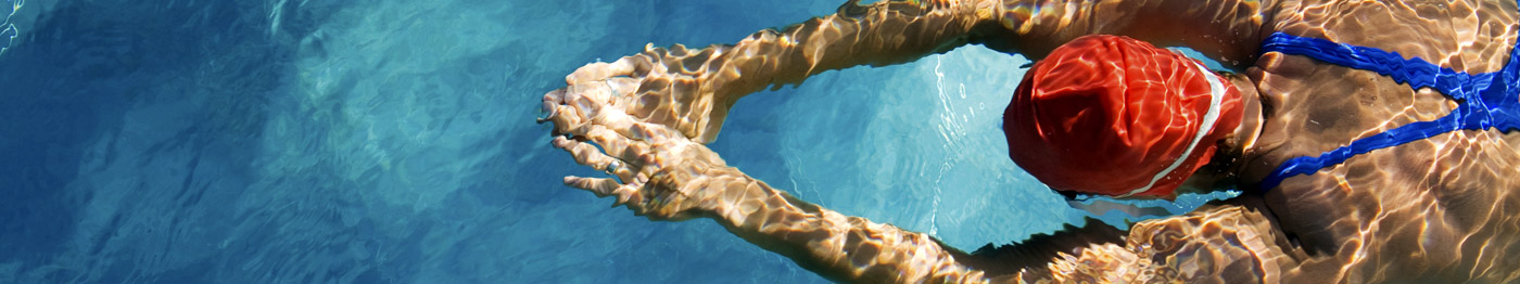
Exercises in a pool can usually be started after two weeks when the wounds have healed. Gentle swimming and cycling on an exercise bicycle should be undertaken after 4 weeks and light weight training may be undertaken after 6 weeks. This will speed up the rehabilitation. Breaststroke swimming should be avoided during this time. The knee should be protected from impact and excessive strain during this time.
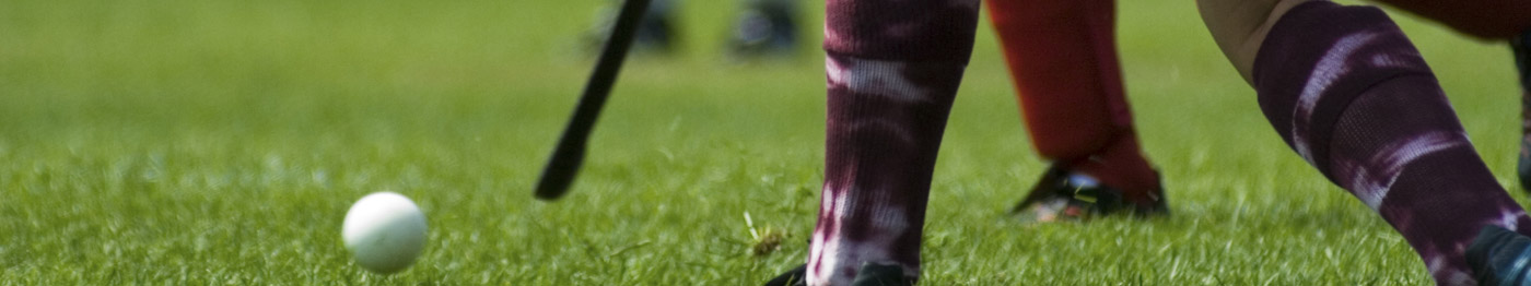
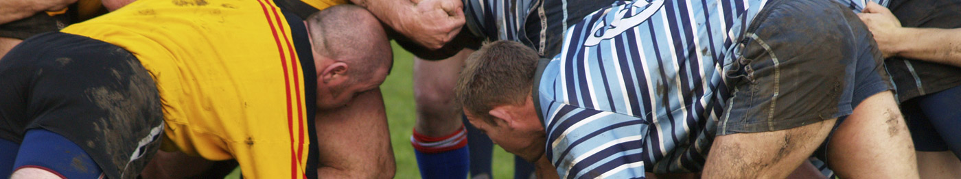
After 3 months normal outdoor cycling, normal swimming, weight training and golf may be undertaken. Gentle jogging on a straight line on a running machine or grass should be performed. After 6 months normal running including rough ground and twisting can be performed. Tennis, badminton and non competitive squash is allowed. Training for soccer, rugby, hockey and basketball may be started with gentle kicking of a ball but tackling should be avoided in recreational athletes at this time. Professional atheletes may wish to return at a slightly earlier time in the rehabiltation process.
After 9 months competitive soccer, rugby, hockey and basketball may be undertaken but the ligament will not reach its full strength for 12 months, so take care. At 12 months full return to sporting activity and skiing is allowed
Author:
DAVID P JOHNSON
MB ChB FRCS FRCS. MD
Consultant Orthopaedic Surgeon
www.Bristol-Knee-Clinic.co.uk
© OrthopaedicsOpinionOnline 2011 www.OrthopaedicOpinionOnline.co.uk
Disclaimer: The views expressed in this article are not necessarily those of Orthopaedic Opinion Online or the author. The information is provided for general background reading only and should not be relied upon for treatment. Advice should always be taken from a registered medical practitioner for individual circumstances and for treatment of any patient in any circumstances. No liability is accepted by Orthopaedic Opinion Online, or the author in respect to the information provided in respect of the content or omission or for any reason or as a result of treatment in individual circumstances. This information is not for use in the USA.