Knee & Sports Injury Clinic
Bristol
Email: boc@orthopaedics.co.uk

Treatments
Browse through a rich range of information to find details of various treatments available for your condition. These include details on the treatment options, medications, physiotherapy and injections. Details are also provided on the range of surgical procedures and the rehabilitation following surgery.
You can also find out more about how to access private treatment when paying for yourself and the statutory quality criteria and success rates of our treatments.
Hover over links below to view summary or click on the link to view full article:
Arthroscopic Knee Surgery: A Patient’s Guide
THE ANATOMY OF THE KNEE
The knee is a complex joint comprising; bony surfaces covered with a smooth articular cartilage layer. In the knee between the principal two bone of the femur and the tibia are two crescentic shaped fibrocartilage pads called “menisci” or “cartilages”. The ligaments that stabilise the knee consist of the collateral ligaments; medial and lateral, lying either side of the knee and the cruciate ligaments, anterior (ACL) and posterior cruciate ligament (PCL), lying within the joint. The fibrous capsule, which surrounds the knee, completes the stability of the joint. Two of the ligaments of the knee are within the joint; these are the anterior and posterior cruciate ligaments. In addition there are two ligaments outside the joint; the medial collateral ligament and the lateral collateral ligament.
EXERCISES AND MEDICATION: PRIOR TO SURGERY
Prior to surgery and post operatively it is important to strengthen the muscles of the leg and to reduce the stiffness. Regular exercises should be undertaken to do this. Static quadriceps exercises consist of tensing the muscle on the front of the thigh whilst the knee is straight. Hold the contraction for 5 to 10 seconds, rest for 5 or 10 seconds and begin again. This should be repeated 10-50 times. Whilst lying on your back the straight leg should be lifted into the air and held for 5 to 10 seconds, then lowered rested for 5 to 10 seconds and repeated 10 to 50 times. If possible these exercises should be undertaken against weight or resistance or in a gym using exercise equipment.
Anti-inflammatory tablets (Indomethacin, Voltarol, Brufen, Naprosyn etc) must be stopped 2 days before surgery. On the morning of surgery patients should fast from midnight and arrive at the hospital at 7.20 am. For afternoon surgery you will be fasted after 8 a.m. Prior to the operation any tablets or medications you take, or allergies you may have to medications, should be brought to the attention of the surgeon. Please notify your surgeon and anaesthetist in advance if you are taking any anti-coagulants (blood thinners), hormone replacement tablets, the Pill or suffer from diabetes or any other significant medical condition.
The anaesthetist and surgeon will see you before surgery. The operation is performed under spinal or general anaesthesia. There will be one or two small incisions in the front of the knee either side of the patellar tendon.
SURGICAL TECHNIQUE
Arthroscopic, telescopic, keyhole or minimally invasive surgery is the technique of performing surgery inside A joint through a telescope without disrupting the surrounding structures. Looking inside the joint with the arthroscope allows the surgeon to look directly at the cartilages, ligaments and the articular surfaces without damaging the joint. The arthroscope is a pencil thin tube containing light fibres and is a means of transmitting a picture of the inside of the joint to a video camera. The knee joint is filled with fluid which allows the surgeon to look around the brightly lit inside of the joint. The arthroscope is inserted through a small incision, about 5mm long either side of; and just below the knee cap (patella). As well as the arthroscope a small metal probe isinserted into the knee to help show if there are any tears in the menisci.
Once extent of the damage to the menisci or other structures in the knee has been determined, small cutting tools are inserted into the joint through the same holes that were used for the arthroscope. With the arthroscope in the joint giving the surgeon a clear view, these small cutting instruments are then used to trim away the damaged part of the meniscus. This is done so that there is once again a smooth surface allowing the joint to roll back and forth without catching or locking. The pieces of meniscus are then removed through the same small holes as the joint is washed out. The fluid is drained out at the end of the procedure, however the knee may feel as if there is a little fluid within it for a few days.
Arthroscopy is performed whilst causing as little disruption to the knee as possible, with a minimal of post-operative pain and with a rapid return of function. Patients can generally return to work in a day or two, to sports in a week or two and to normal thereafter. The results and speed of recovery is very much dependant on the nature of the damage to the knee and the extent of surgery undertaken and may as a consequence vary from this norm.
In the case of a tear of the menisci, or “cartilages” of the knee the smooth rolling of the joint surfaces over the meniscus is prevented as the torn meniscal flap gets caught between the bones of the tibia and the femur. The knee is usually painful, particularly on twisting, swollen and cannot be fully straightened. Arthroscopic partial meniscectomy is undertaken to remove the torn piece of meniscus to relieve the pain and swelling, to allow full movement and to prevent further damage to the knee. The amount of surgery required to remove this torn portion of meniscus will not be known for certain until the surgeon can see the damage through the arthroscope. However the aim is always to remove as little as the tear allows.
In some cases the meniscus may be able to be repaired. This is where the tear is a “peripheral bucket handle” type, is fairly recent in a patient who usually is under 40 years of age. In this situation the surgeon may be able to suture or stitch the tear edges together through the arthroscope so that it may heal after a period of six weeks. Where possible this may be advantageous in preserving the meniscus and avoiding it’s removal. This minimises any potential for excessive knee wear in the future.
The anaesthetist will see you before surgery. The operation is performed under spinal or general anaesthesia. There will be one or two small incisions in the front of the knee either side of the patellar tendon. A steristrip or a single suture is used to close these wounds.
Following the operation you usually wake up without or with only modest pain because local anaesthetic is inserted into the joint at the end of the procedure. This may wear off after 4 – 6 hours and the tablets to relieve any paid and anti-inflammatory medication is usually prescribed. By the following morning the leg is usually comfortable whilst taking only regular anti-inflammatory medication. Generally only a few hours after your operation you will be able to get around walking without too much difficulty and without crutches. Most arthroscopic surgery patients can be discharged form hospital later the same day as a “Day Case”. Sometimes when extensive surgery is performed or operations preformed simultaneously on both legs patients are kept in hospital overnight. After patients go home, they should keep bending and lifting the knee in order to strengthen the muscles and regain movement.
If the knee becomes more swollen over the first week or more painful than during the first day, please return to your general practitioner for early review. Normally you will be reviewed at the hospital by your surgeon after 2 or 3 weeks.
WOUND DRESSING AND SUTURES
The dressings should be removed after 5 days and the wound inspected. If there is any excessive redness or infection patients should return to the GP or the clinic. If the wounds have been closed with steristrips these may be removed at that time. If sutures were used then the dressings should be maintained for 10 days, following which you should return to the GP’s clinic to have the stitches removed. A tubigrip elasticated support should be worn for 7 – 10 days. The wound if dry and the sutures have been removed or dissolved then the wound can be washed, and pool exercises can be begun. If the wound becomes red, inflamed or infected then patients should return to see your surgeon.
MEDICATION
If you do not suffer from gastric irritation, you should take anti-inflammatory tablets for two or three weeks to settle down the inflammation and swelling in the knee. The anti-inflammatory tablets should be taken after eating. If nausea, vomiting or abdominal pain develops you should reduce the dosage. If despite reducing the dosage gastric irritation continues then the tablets should be stopped and you should contact your general practitioner.
PHYSIOTHERAPY
Prior to surgery quadriceps exercises should be performed to strengthen the knee. These should also be performed immediately after your operation on waking. A physiotherapist should visit you after the operation to assist you to stand and start your mobilisation. The physiotherapist will advise you on the exercise program and supervise your quadriceps exercises. Crutches or sticks are not usually necessary. The physiotherapist will also assist you to try bending the knee a little. When you are comfortable and able to walk, and the nurses and physiotherapist are happy with your progress, you may be collected and taken home. This is usually 5 pm for morning cases and 8 pm for afternoon cases.
The exercise program of quadriceps exercises should be vigorously continued at home. Approximately 5 minutes each hour will be ample. After 3-5 days more knee flexion should be possible. An out patient physiotherapy session should be arranged during this period. After 7 days knee bending should begin to return to normal. Resisted exercises and light weight training may be undertaken if desired, this would be beneficial. The progress is variable, so do not worry if your progress is a little slow at this stage.
RETURN TO WORK AND SPORT
If your job is sedentary and mostly sitting you may wish to return after only 1 or 2 days. If your job is physically demanding and requires standing or walking for most of the day, your return to work may take 1-2 weeks. Driving can usually be performed for short periods after 2-5 days providing that the knee is pain free and you are able to control the car with foot pedals and make an emergency stop.
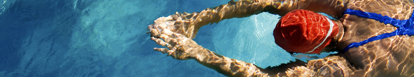
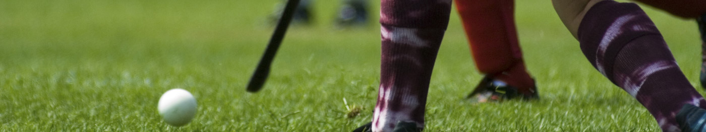
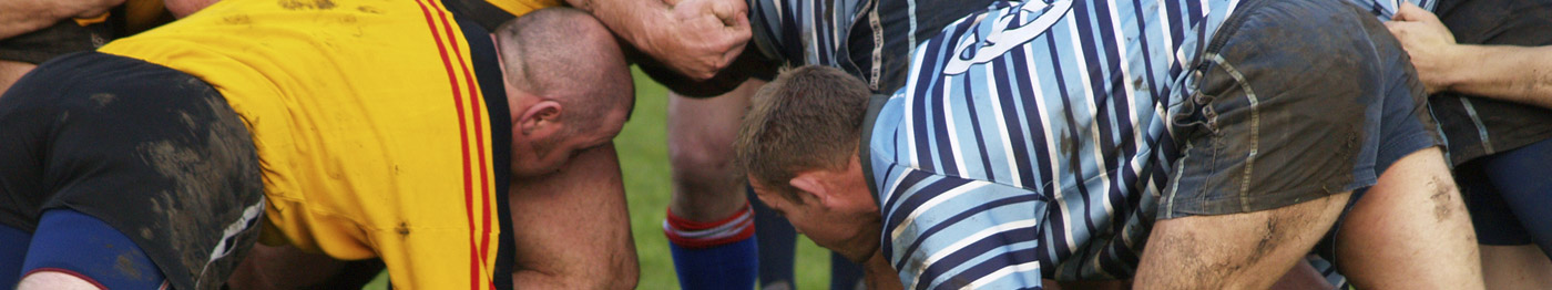
Light cycling, swimming, weight training or jogging may be undertaken after 7 days. This will speed up the rehabilitation and should be performed prior to undertaking more vigorous sports. Return to running, soccer, rugby and squash is usually after 1-4 weeks depending on individual progress.
Please visit www.orthopaedics.co.uk for more information.
DAVID P JOHNSON
MB ChB FRCS FRCS. MD
Spire Glen Hospital, Redland Hill, Bristol BS6 6HW. UK
Appointments: 0117 970 6655
E-Mail: boc@orthopaedics.co.uk
Web site: www.orthopaedics.co.uk
© OrthopaedicsOpinionOnline 2011 www.OrthopaedicOpinionOnline.co.uk
Full text pdf
Disclaimer: The views expressed in this article are not necessarily those of Orthopaedic Opinion Online or the author. The information is provided for general background reading only and should not be relied upon for treatment. Advice should always be taken from a registered medical practitioner for individual circumstances and for treatment of any patient in any circumstances. No liability is accepted by Orthopaedic Opinion Online, or the author in respect to the information provided in respect of the content or omission or for any reason or as a result of treatment in individual circumstances. This information is not for use in the USA.