Knee & Sports Injury Clinic
Bristol
Email: boc@orthopaedics.co.uk

Treatments
Browse through a rich range of information to find details of various treatments available for your condition. These include details on the treatment options, medications, physiotherapy and injections. Details are also provided on the range of surgical procedures and the rehabilitation following surgery.
You can also find out more about how to access private treatment when paying for yourself and the statutory quality criteria and success rates of our treatments.
Hover over links below to view summary or click on the link to view full article:
Rationale, Indication, Technique and Results of Arthroscopic Surgery for Patellar Tendonitis.
Nottingham International Knee Meeting, October 2000.
Arcus Sportsklinic, Fortzheim, Germany, November 2000.
Abstract: A short review of the development and use of a new surgical procedure for the treatment of patellar tendonitis.
Patella Tendonitis
Patellar tendonitis is a very common condition which is poorly understood, poorly treated and often troublesome. It is commonly known as “Jumpers Knee” as described by Blazina in 1973. Patella tendonitis is often confused with other conditions affecting the patella tendon. Specifically this condition affects the superior central and posterior aspect of the patella tendon in close proximity to the inferior pole of the patella. It commonly causes pain during and after activities, particularly repetitive activities, descending stairs or jumping.
There are many misconceptions about patella tendonitis, such as the belief that the condition spontaneously improves and that rest is all that is necessary to effect a cure, that physiotherapy is commonly successful, that surgery is never necessary, and that an arthroscopic treatment for this condition would be dangerous.
There are many intrinsic factors which are known to pre-dispose to the development of patella tendonitis. These include limb mal-alignment, leg length discrepancy, foot hyper-pronation, muscle imbalance, fatigue and poor flexibility, particularly of the hamstring muscles. Other associated factors include poor vascularity, diabetes and gout. The condition is more common in females. The condition does not affect juvenile immature skeletons but does affect young athletes and ageing athletes in the 30 to 40 age group. Juvenile patients suffer from Sinding-Larsen-Johanssen’s disease of an apopysitis of the inferior patellar pole; a different condition. Patellar tendonitis is also increased in frequency where there is a history of previous injuries, particularly trauma to the anterior aspect of the knee or a direct blow to the inferior pole of the patella.
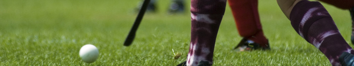
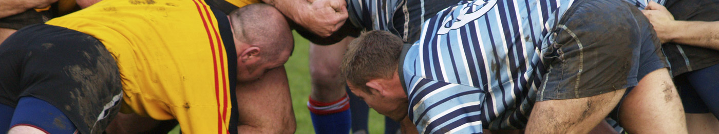
Extrinsic factors may also play a part. These include excessive load, such as jumping, basketball or volleyball. Excessive driving or flying (ie piloting) in a seated position. Jogging on hills or stairs and step aerobics may all be problematical. Using a poor position or technique during jumping or running may also precipitate the condition. Environmental factors such as the surface or floor used for exercise and poor equipment, particularly shoes, can play a part. Sporting rules are important, as in Netball when landing an instant stop is necessary.
Treatment of Patellar Tendonitis
There are further misconceptions about treatment of this troublesome condition. There is no evidence that many commonly used treatments are at all effective. These include, rest, immobilisation in a plaster cast, massage and frictions, muscle strengthening and VMO exercises, McConnell taping, and anti-inflammatory gel applied locally. Other modalities have proved to be helpful but have not undergone proper audit. These include altering of driving position, avoiding road running and hill running, adjusting the cycling position, use of orthotics or a pre-patellar splint, and hamstring stretching.
There are a few treatments that have been proved to be effective and these include all anti-inflammatory medication, eccentric quadriceps physiotherapy as described by Standish and the various surgical treatments which have been reported.
It has been shown that steroid injection into the tendon may be dangerous and is associated with tendon degeneration and disastrous rupture. Steroid effects include a local anti-inflammatory effect and pain relief but a consequence of this may be tendon degeneration. Such injections should be avoided, exceptionally if they are used it must be after counselling of the patient and the injection should not be placed in the tendon itself, but in the fat pad on the posterior central and superior aspect of the tendon. Non steroidal anti-inflammatory medication is an essential part of the non-operative treatment and should be used for a period of 3-6 weeks in order to gauge it’s effect.
Generally describing the management of this condition. The initial management should include alteration of the physical activities of the patient. This includes running which may need to be curtailed, certainly running on hills or hard surfaces should be avoided. Cycling should be modified to avoid hills and to use a high saddle position. Jumping and repetitive activities should be avoided. Analysis of shoe wear should also be undertaken. Alteration of the driving position and avoidance of a stiff clutch may prove to be helpful. Drivers of four-wheeled drive vehicles are prone to develop this condition. Patients should also undertake a rigorous stretching programme particularly of the hamstring, calf muscles and lumbar spine.
Physiotherapy
Physiotherapy management includes supervision and encouragement of the stretching programme. Massage is commonly used but it’s effect is unproven. Ice may have a useful pain relief and anti-inflammatory effect. Static strengthening of the quadriceps is commonly undertaken, however, repetitive eccentric and concentric extension exercises against resistance, particularly open chain exercises, may be counterproductive. Orthotics should be prescribed as necessary and some patients may have symptomatic relief from the use of a pre-patellar strap. Standish in 1986 reported the results of physiotherapy for this condition. After thorough completion of a treatment programme patients were reviewed after one year. Only 44% could be said to be excellent and symptom free, a further 43% had improved but were still troubled by the condition. Nine per cent had no response from physiotherapy and 2% were worse. The treatment described by Standish included, in addition to stretching eccentric quadriceps dips in what was a fairly extensive and prolonged treatment protocol.
Therefore a significant proportion of patients with this syndrome will not be improved by non-operative means and will present with continuing symptoms. The indications for surgery are, pain in the region of the inferior pole of the patella, restrictive to the patients activities which has proven resistant to non-operative therapy. This is generally reserved for the grade 2 or 3 symptoms described by Blazina or persistent grade 1 symptoms in serious athletes.
Historically many different treatment modalities have been described for this condition, previously the most popular treatment had been excision of the degenerative nodule which can often be found on the posterior aspect of the patella tendon. This was described by Orava, Osterbach and Hurme in 1986 and Albright in 1990. Orava followed this treatment by a long period of immobilisation and reported 67% good or excellent results following his treatment.
Perugia et al, in 1981 and Albright in 1990 described the details of the procedure, this included a longitudinal incision through the superior central portion of the tendon. Excision of the degenerative nodule on the posterior aspect of the tendon, drilling of the inferior pole of the patellar or excision of the inferior pole of the patella and a period of immobilisation for 4 – 8 weeks, this resulted in a return to sports after 3 – 4 months. Detachment of the central posterior portion of the patella tendon from the inferior pole was described as an additional procedure further to excision of the degenerative nodule. Blazena, Kirland and Jobe in 1973 (Orthop Clin. North America) championed this first. Other notable surgeons described the same procedure, including Martens, Wouters and Burssens (Acta Orthopaedica Scan 1982) and Orava Osterbach and Hurme (British Journal Sports Med 1986). Other procedures described for this condition include drilling or even excision of the inferior pole of the patella. This was suggested by Smilie in his textbooks in 1974, and Orava, Osterbach and Hurme in the British Journal of Sports Medicine 1986.
Generally all these treatments had varying levels of success at between 60% and 93% of cases. The differences in the surgical technique was completely subjective and arbitrary without any clear rational or justification, therefore the surgeon was left without any guidance as to whether simple excision of the degenerative nodule was sufficient or, whether a curettage drilling excision of the inferior pole was necessary or, even as described by some authors reattachment of the patella tendon to inferior pole with sutures.
In order to determine which of the surgical treatments would be preferential, some ten years ago I started project of research to analyse and investigate this condition in order to determine the aetiology, pathology and anatomy, to define the appropriate investigations and determine the optimum treatment protocol.
Anatomy
The first aspect I studied was an analysis of the anatomy of the patellar tendon. This has recently been published in the Journal of the European Society of Sports Surgery, Knees Surgery and Arthroscopy by my research fellow Mr. Oreste Basso. We were able to show that the anatomy described in the textbooks and surgical papers was incorrectly described. The patella tendon inserts to the margin of the inferior pole of the patella and the anterior surface. Between the inferior edge of the articular surface and the inferior pole there was a “bare area” of patella covered only by fat pad. Thus the inflammatory nodule seen clinically on the posterior aspect of the tendon is a swelling and out-pouching of the tendon, rather than an area of tendon which has become detached from the posterior aspect of the inferior pole of the patella.
MRI
Analysis of the MRI appearances and morphology of the patella was then undertaken, as reported by myself and Charles Wakerley in the British Journal of Bone and Joint in 1996. We were able to show that the characteristic lesion is an expansion of the patella tendon in the area where the tendon inserts into the inferior pole of the patella. The lesion is in the central posterior and upper part of the tendon and always associated with the inferior pole. The MRIappearances seem to suggest that the tendon inserts into the posterior bare area of the inferior pole, however, from the anatomical studies this is known to represent a swelling of the tendon, inflammation and a high MRI signal in this area.
Pathology of Patellar Tendonitis
When analysing the morphology in this condition from the MRI measurements it was noted that the inferior pole is not significantly elongated in these patients, however, sporadically some patients do present with a long inferior patella pole, which is thought to predispose to this condition. Patients did have an elongated patella tendon and a degree of patella alta. This was without any degree of patella subluxation. We demonstrated that patients were of above average height. In our initial patient study we noted that patients had pre-disposing activity factors such as long distance driving, particularly four-wheeled drive vehicles, flying small aircraft. Sports activities were common but more particularly jogging as opposed to jumping activities was a factor, and most patients in our study population had a significant degree of hamstring tightness.
I also analysed the histology of this condition. Puddhu had initially described the histology as showing synovial proliferation, fibrinoid necrosis, mucoid degeneration and an increased vascularity in the tendon. This was assumed, at the time, to represent microscopic tears due to a stress overload, which in some cases accounted for a rupture of the patella tendon. However, although we were able to show some small degree of mucoid degeneration in the central portion of the superior tendon, the increased vascularity was confined to the posterior aspect of the tendon. Haemosiderin deposition occurred in this area, indicative of repetitive trauma. There were no microscopic tears in the tendon. The histology, therefore, suggested a tendonosis rather than a tendonitis and there was no evidence that this was a stress overload failure, rather a repetitive trauma compression and or impingement.
Surgery for Patellar Tendonitis
Considering these results and the reported clinical success from release of the posterior central portion of the patella tendon made me wonder whether this condition was indeed a stress overload failure. If it had been then following surgical treatment where the tendon is weakened, detached or excised one would expect the condition to become more significant rather than settle as in the majority of cases. It was further noted that the condition in jumping athletes occurs not on the stress take off phase of a jump, but on the landing, deceleration, eccentric quadriceps phase. This was also the knee position adopted when driving and flying which was the precipitating factor in a significant proportion of our patients. From these results I formulated and proposed the impingement theory of the aetiology of patella tendonitis, which has subsequently been generally adopted and accepted. This suggests that the condition is produced by a repetitive compression of a posterior superior central portion of the patella tendon by the inferior pole of the patella. This occurs in flexion and eccentric motion such as landing from a jump where the quadriceps is under tension and the quadriceps and patella tendon effect is to decelerate the patella and femur. The point of maximum flexion is where the tendon curves around the inferior pole of the patella. This would explain why the condition also affects patients when driving, flying or jogging on hills. It further explains the good surgical results following detachment of the posterior superior central fibres of the tendon from the inferior pole and excision of the inferior pole of the patella. This would also be entirely compatible with the histology that was found.
We have further investigated this with the help of Professor Andrew Amis at the Bio-mechanics department of Imperial College London. We have undertaken various bio-mechanical studies which have been variously reported by myself, Andrew Amies and Oreste Basso in the literature. This has confirmed that the posterior central fibres of the patella tendon are the shortest fibres and those subjected to greatest strain during loading of the knee in flexion. Further the compression of the tendon by the inferior pole of the patella is greatest in the position of 30 degrees to 60 degrees of knee flexion: the position of knee flexion in which symptoms occur. We have further shown that excision of the inferior pole is the most effective surgical technique for decompression of this area in the laboratory.
The conclusion of my initial research, therefore, is the embodied by the formulation of the impingement theory of the pathogenesis of patella tendonitis. This theory is able to explain the various factors associated with the condition such as the eccentric activity, repetitive activity, hamstring tightness, the excessive patient height, long patella tendon and inferior pole lengthening commonly associated with this condition. It also explains the characteristic posterior superior and central area for this condition, the tendonosis, , the haemosiderin deposition on the posterior aspect of the tendon, the biomechanical properties of the tendon and the success of surgical release of the tendon from the inferior pole and or excision of the inferior patella pole.
In considering the applicability of the various surgical techniques described. If the condition is indeed a compression of the inferior pole against the posterior aspect of the tendon, then as in the analogous situation of rotator cuff syndrome in the shoulder, excision of the inferior patella pole would be the most appropriate surgical technique. This would be more logical than drilling or curettage of the inferior pole, trefining of the tendon, reattachment of the tendon, or indeed release of the central portion of the tendon. It also explains why simple excision of the nodule may well be ineffective.
Arthroscopic Debridement Procedure for Patellar Tendonitis
I designed, therefore, a surgical technique whereby the inferior pole of the patella could be excised but undertook this using minimal invasive techniques for the first time by undertaking this via the arthroscope. This was expected to be advantageous in allowing a thorough examination of the knee, and simultaneously be minimally invasive, and have the associated advantages of a rapid rehabilitation and the other usual advantages of minimal access surgery.
I undertook, therefore, the first arthroscopic decompression for patella tendonitis in 1992. The surgical technique has only been slightly modified since the initial procedure.
The technique involves a standard arthroscopy using a standard 30 degree arthroscope. This utilises the antrerio-medial and anterio-lateral portals. I initially also utilised the superio-lateral and superio-medical portals to gain a wider view and appreciation of the infra-patellar fat pad and used a 70 degree arthroscope. With increasing experience this has proven unnecessary. The arthroscopic appearances of the patellar articular surface and the fat pad is usually entirely normal. The fat pad covering the non-articular “bare area” of the inferior pole of the patella is reflected using arthroscopic shaver 4.5 mm in diameter. The opening on the blade is kept towards the bare area of the patella. It is very important to expose the whole of the anterior surface and margin of the inferior pole. The position of the shaver can be checked by use of a percutaneous needle placed at intervals into the knee at the position of the tip of the inferior pole. The patella tendon is then elevated from the anterior surface of the inferior pole for a distance of approximately 3 mm. This is undertaken keeping the opening of the shaver blade in close contact with the inferior pole and by a stroking action, elevate the tendon fibres from the inferior pole. This is assisted by a surgical assistant pressing with a single finger over the lower part of the patella, delivering the inferior pole into the knee and into the view of the arthroscope. Initially a 70 degree arthroscope was used to improve the visualisation of this, however, with experience this is unnecessary and unhelpful.
Once the inferior pole is exposed the tip of the inferior patella pole is removed. This is undertaken after confirmation of the exact location of the inferior patella pole by a percutaneous needle. A 4.5 mm spherical arthroscopic burr is used to remove some 3-5 mm form the inferior pole. This usually involves a quarter to a third of the non-articular inferior pole of the patella. The resection must be extended anteriorly to the anterior cortex, through the whole thickness of the patella. This avoids leaving an anterior lip, which may continue to compress the tendon. Switching to an antero-medical viewing portal and an anterior-lateral surgical portal facilitates excision of the inferior pole to a depth of 3-5 mm and for a distance of 5 – 7 mm either side of the inferior pole. It is very important to avoid damage to the articular surface and to the patella tendon itself or to the patellar tendon. As the majority of the tendon fibres insert to the anterior surface of the patella itself these fibres can be elevated and the inferior pole excised without significant damage to the tendon itself. With experience the procedure can comfortably be undertaken in 20 – 30 minutes, however, initially considerable expertise, time and patience is necessary to gain adequate visualisation and to undertake the procedure safely and successfully.
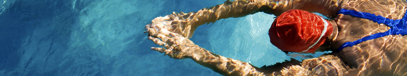
The post-operative rehabilitation includes initial full weight bearing, discharge as a day case from hospital, anti-inflammatory medication for 3 – 6 weeks. The patient returns to the activities of daily living and driving in 3 to 5 days. Range of motion exercises, regular ice-ing and control of the swelling is undertaken in the first 3 weeks. Formal physiotherapy and strengthening exercises using static quadriceps activities are started at three weeks. Light sport such as static cycling and swimming can be undertaken at 6 weeks and return to jogging and sports training is usually undertaken between 6 and 12 weeks after surgery. Patients should be warned that the recovery from this procedure is not the same as from an arthroscopic meniscectomy, and return to activity is not undertaken as rapidly.
My experience with this procedure was reported in a minimum 5 year review of 20 patients. There was an 85% follow up of 17 patients, 15 male, 2 female for a range of 2.6 to 6.4 years. Prior to surgery 90% of patients had Grade 2 or 3 patella tendonitis. At 2 years post-operatively 80% had no symptoms and 20% had Grade 1 symptoms, and at 5 years 94% had no symptoms and 6% (one patient ) had Grade 1 symptoms. In no case was re-operation necessary.
There tended to be a continued improvement in the symptoms between the 2nd and 5th year review. In this period no case deteriorated and the good and fair results improved. The single poor result did not seek further treatment for this period and did not improve. I reported, therefore, the advantages of this procedure of arthroscopic decompression for patella tendonitis over the traditional open methods. I brought attention to the fact that the inflammatory nodule is a secondary feature and does not necessarily need excision. I described the practical advantages of excision of the inferior pole over the other surgical techniques. Arthroscopic decompression allows for a simultaneous arthroscopy, day case surgery, early return to normal activities and sports and an increased chance of a successful outcome. The outcome of 94% good long term results is better than that previously reported from other techniques in the literature.
The disadvantages of this technique are that it requires experienced arthroscopists, patients do have some discomfort for 1 – 3 weeks and may have some swelling for 1 – 2 weeks. Return to sport is not instantaneous and is delayed between 6 – 12 weeks. The reported failure rate in the initial study was 6% although this was the first patient on which this procedure was used and subsequent results have been without such failures.
My total experience with this technique was analysed several years ago this included the first 44 patients with a 1 – 6 year follow-up. In this group 93% had good or excellent results and 3 patients had fair or poor results. Once again there were no complications and no re-operations proved necessary. I also reported the first 9 cases where this technique had been undertaken following failed previous open surgery undertaken by others. The 9 revision cases had had a total of 17 previous operations, they were followed up with a 1 – 2 year follow-up. Of the 9 cases 7 were good and symptom free, 2 patients continued to have mild symptoms but had been improved by the revision procedures. There have been no complications.
Conclusions
I was therefore able to conclude that the initial five year review of this technique of arthroscopic decompression for patella tendonitis proved to be technically feasible, safe with as yet no significant complications, successful in the long term in 94% of patients and could be undertaken routinely as a daycase and was associated with rapid rehabilitation and return to sport.
Further this success following excision of the inferior pole of the patella and decompression of the patella tendon, without excision of the degenerative nodule supported the theory, that this condition is a result of an impingement or compression of the inferior pole upon the posterior aspect of the tendon in flexion rather than a failure in tension.
David P. Johnson MD.
Consultant Orthopaedic Surgeon
Spire Hospital Bristol,
Redland Hill, Bristol, BS6 6UT
boc@orthopaedics.co.uk
www.Bristol-Knee-Clinic.co.uk
November 2000.
© OrthopaedicsOpinionOnline 2011 www.OrthopaedicOpinionOnline.co.uk
Full text pdf
Disclaimer: The views expressed in this article are not necessarily those of Orthopaedic Opinion Online or the author. The information is provided for general background reading only and should not be relied upon for treatment. Advice should always be taken from a registered medical practitioner for individual circumstances and for treatment of any patient in any circumstances. No liability is accepted by Orthopaedic Opinion Online, or the author in respect to the information provided in respect of the content or omission or for any reason or as a result of treatment in individual circumstances. This information is not for use in the USA.